616-785-CORE (2673) / info@coreimaging.com

Read this case study for tips on performing scans on obese pets.

Case Study: Verde Veterinary Hospital

https://21907686.fs1.hubspotusercontent-na1.net/hubfs/21907686/Core_Imaging_September_2023/images/AdobeStock_164526817.jpg

Dr. Audrey Yank used CoreVu on her Piloter ultrasound system to complete an abdominal scan.

The Study:

  • Images on obese animals are incredibly difficult
  • Slight adjustments to the system and suggesting a different transducer approach was the key
Case Study: Verde Veterinary Hospital

Results of Study

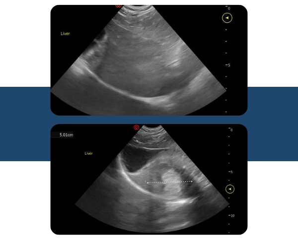
  1. The liver is significantly heterogenous and has signification decreased vascular conspicuity.
  2. The liver lobe margins are irregular and blunted. The overall size is subjectively enlarged.
  3. Incidental cysts are present.
  4. A large target lesion approximating 5 cm in diameter is identified at the cranial ventral margin. The center of this lesion is hyperechoic.
  5. Acoustic shadows are forming.

The sonographers that support CoreVu help to break down the scan in an organized fashion, following a protocol to visualize all the abdominal organs. CoreVu Scan Assist gives you the ability to have a sonographer instruct you through your entire exam. This technology will help you acquire diagnostic images to be then read by our board-certified radiologist.


Previous

Case Study: Deer Creek Animal Hospital

Next

Top 3 Ways Virtual Support is Making Ultrasound a Must-have for Every Practice